CASE REPORT
Involvement of jawbones as a radiographic sign in multiple myeloma – case series reports
1
Student Research Group, Department of Dental and Maxillofacial Radiodiagnostics, Medical University, Lublin, Poland
2
Department of Dental and Maxillofacial Radiodiagnostics, Medical University, Lublin, Poland
Corresponding author
Emanuela Bis
Student Research Group, radiograph. Department of Dental and Maxillofacial Radiodiagnostics, Medical University, Chodźki 6, 20–093, Lublin, Poland
Student Research Group, radiograph. Department of Dental and Maxillofacial Radiodiagnostics, Medical University, Chodźki 6, 20–093, Lublin, Poland
J Pre Clin Clin Res. 2023;17(4):242-244
KEYWORDS
TOPICS
ABSTRACT
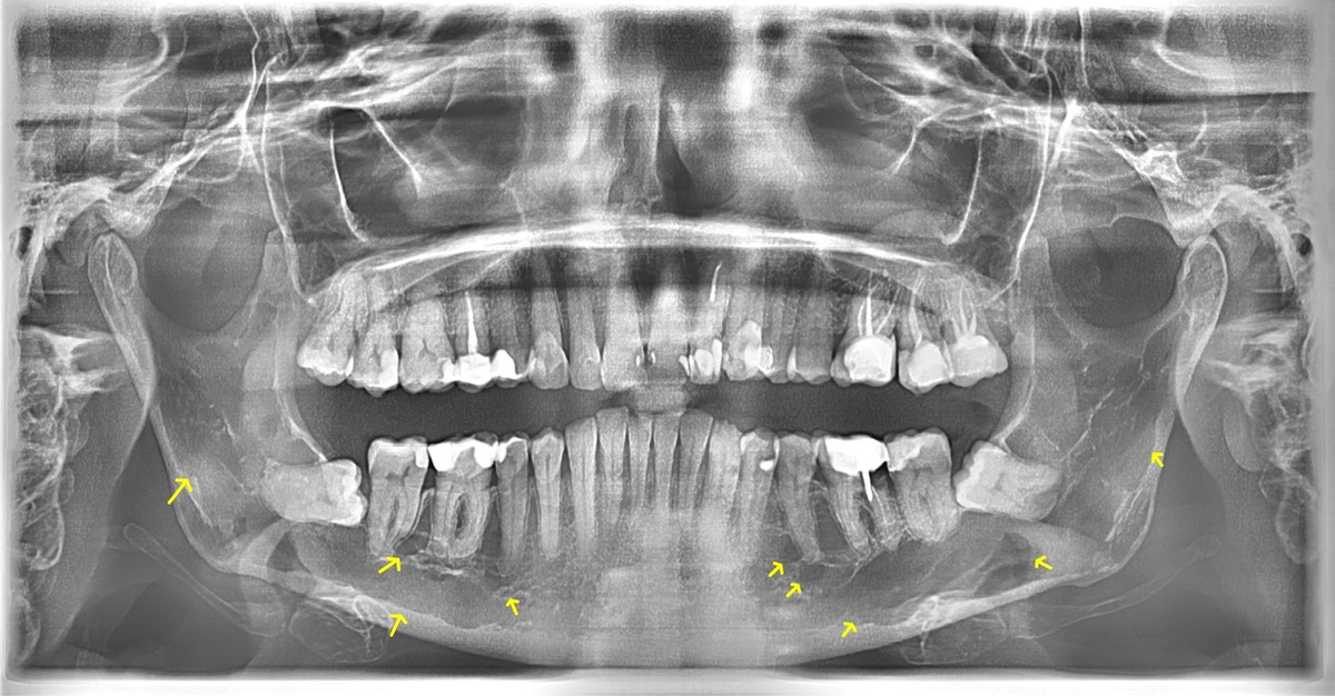
Multiple myeloma is the most common bone tumour characterised by the uncontrolled proliferation of malignant plasma cells. The study presents two case reports of patients with multiple myeloma with manifestation in the maxillofacial region, visualised with the use of a panoramic radiograph. The patients were admitted to hospital due to back pain caused by pathological changes in the vertebral column, including low-energy fractures. The treatment involved a combination of radiotherapy and chemotherapy. Patients were referred for panoramic X-ray and oral cavity sanitation, which revealed an increase in bone mineral loss and the presence of multiple osteolytic lesions. Similar radiological findings were observed in the jaws of both patients. Radiography, including dental panoramic radiographs, plays a crucial role in the detection of osteolytic lesions associated with multiple myeloma. Dentists, during routine check-ups, can be the first to detect osteoporotic changes in the bones.
Bis E, Piskórz M, Różyło-Kalinowska I. Involvement of jawbones as a radiographic sign in multiple myeloma – case series reports. J Pre-Clin
Clin Res. 2023; 17(4): 242–244. doi: 10.26444/jpccr/175609
REFERENCES (6)
1.
Viganò L, Bellini D, Caruso F, et al. Multiple Myeloma – Oral Radiological Evidences. World Cancer Res J. 2021;8:1–9.
2.
Kosmala A, Bley T, Petritsch B. Imaging of Multiple Myeloma. Rofo. 2019;191(9):805–16.
3.
Akyol R, Şirin Sarıbal G, Amuk M. Evaluation of mandibular bone changes in multiple myeloma patients on dental panoramic radiographs. Oral Radiol. 2022;38(4):575–85. https://doi.org/10.1007/s11282...- 00590-6.
4.
Faria KM, Brandão TB, Silva WG, et al. Panoramic and skull imaging may aid in the identification of multiple myeloma lesions. Med Oral Patol Oral y Cir Bucal. 2018;23(1):e38–47.
5.
Lecouvet FE, Vekemans M-C, Van Den Berghe T, et al. Imaging of treatment response and minimal residual disease in multiple myeloma: state of the art WB-MRI and PET/CT. Skeletal Radiol. 2022;51(1):59–80.
6.
Gomez CK, Schiffman SR, Bhatt AA. Radiological review of skull lesions. Insights Imaging. 2018;9(5):857–82.
We process personal data collected when visiting the website. The function of obtaining information about users and their behavior is carried out by voluntarily entered information in forms and saving cookies in end devices. Data, including cookies, are used to provide services, improve the user experience and to analyze the traffic in accordance with the Privacy policy. Data are also collected and processed by Google Analytics tool (more).
You can change cookies settings in your browser. Restricted use of cookies in the browser configuration may affect some functionalities of the website.
You can change cookies settings in your browser. Restricted use of cookies in the browser configuration may affect some functionalities of the website.


